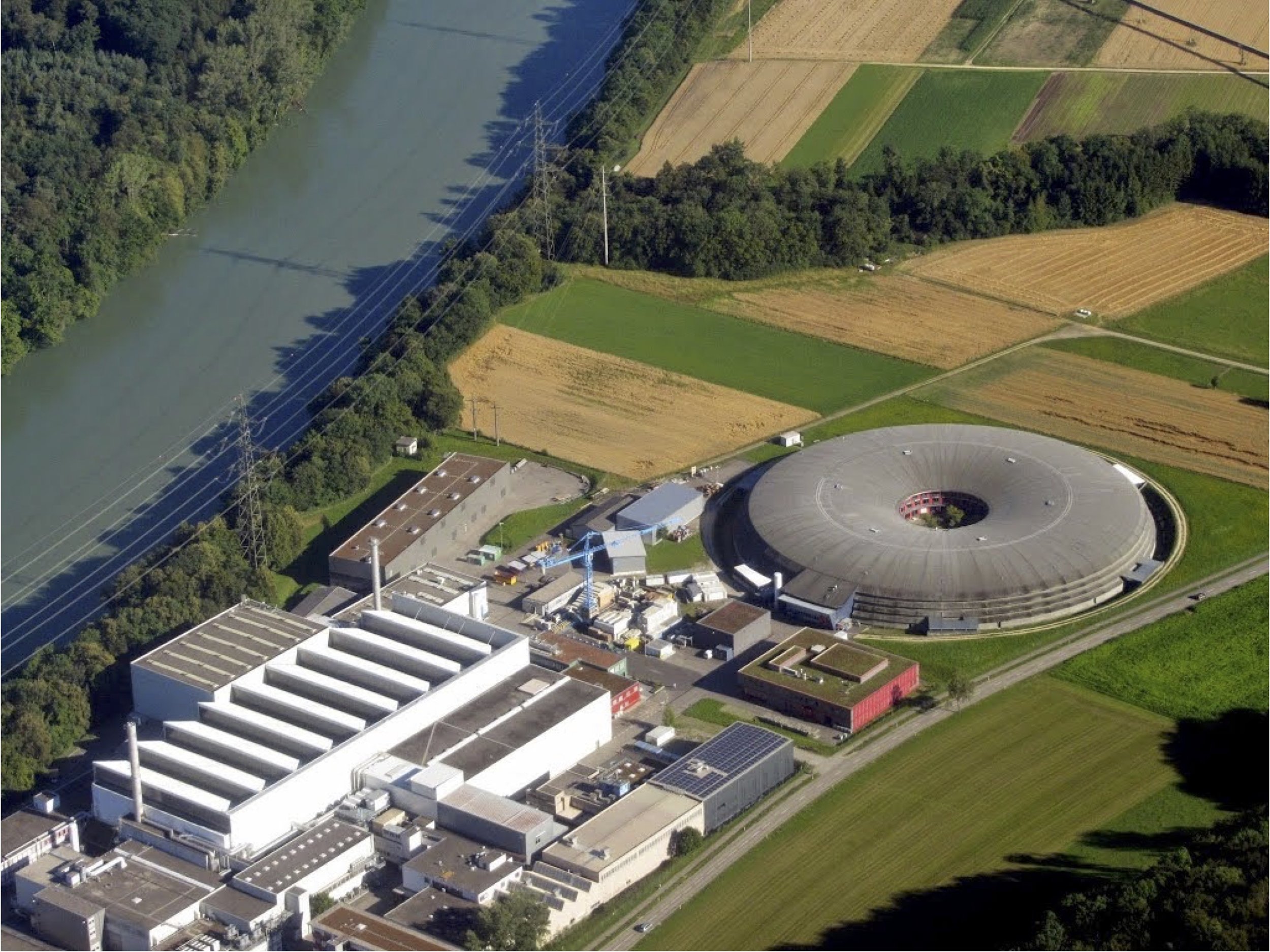
Swiss Light Source Synchrotron
Detailed 3D reconstructions of fossils can be produced via synchrotron imaging. In 2015, I traveled to Zurich, Switzerland, to image blastoids using the Swiss Light Source Synchrotron. Imran Rahman (Museum of Natural History, London) had received a grant of 36 hours of synchrotron time - 36 continuous hours. So, my research assistants, Lyndsie White and Bonnie Nguyen, and I joined Imran, Colin Sumrall and Jen Bauer (University of Tennessee), and Samuel Zamora, an echinoderm worker from Spain. We divided into two groups and worked alternating 8 hour shifts around the clock. Bonnie, Lyndsie and I were running the Tomcat beam line on the Midnight to 8 AM shift on 9/11/2015 when the line went down at 4:50 AM. We did an emergency reboot (in Linux and not for the faint of heart) and got the thing going again.
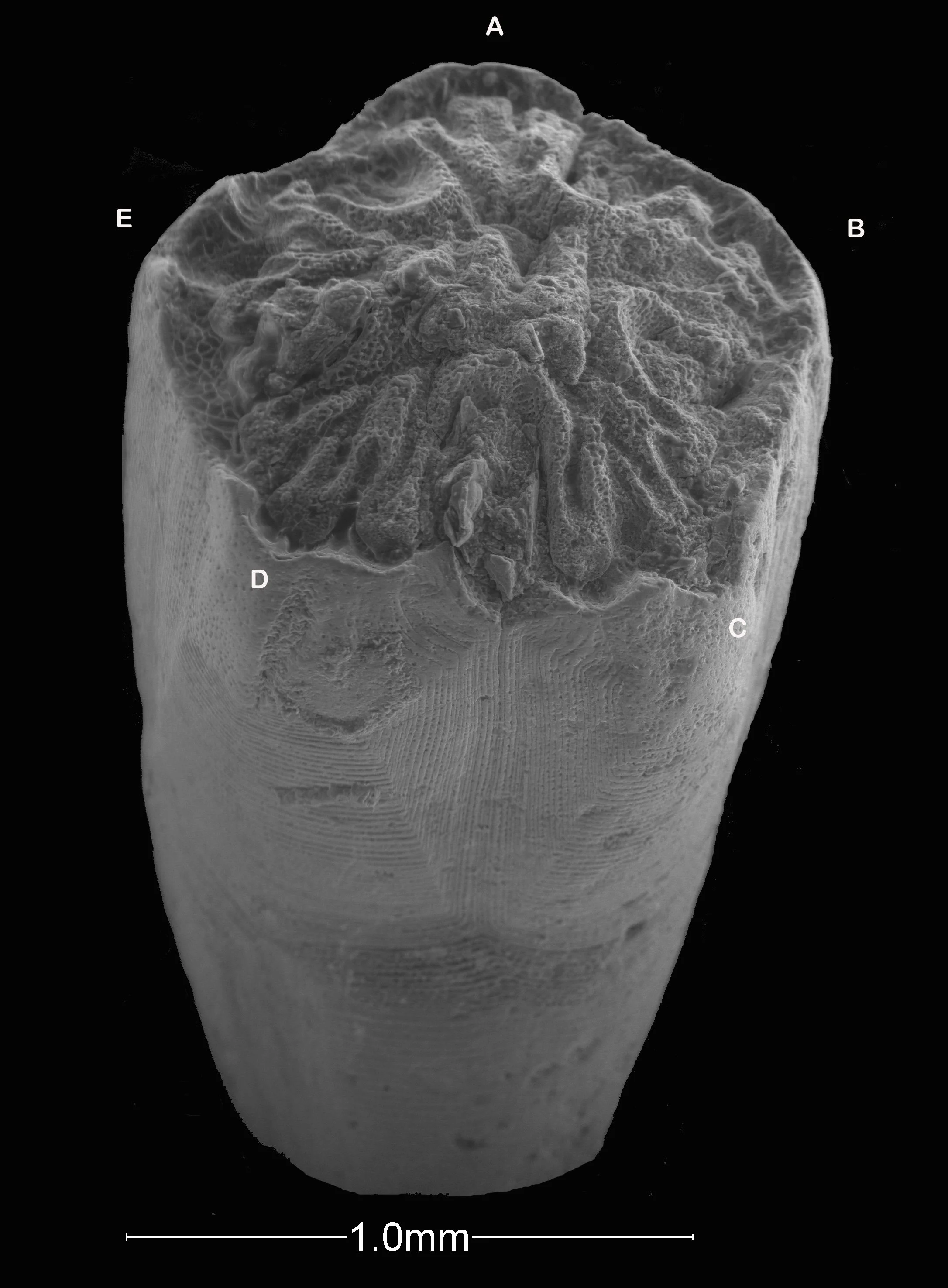
Our most interesting specimen was a 1 mm long larval blastoid that we collected from China in 2003. The specimen (top image) was found by Sara Marcus while looking for other microscopic fossils. Using “propagation-based phase-contrast synchrotron radiation X-ray tomographic microscopy” we were able to identify the gut of the 300,000,000 year old fossil. The first video runs through the slices from top to bottom. Bonnie digitally reconstructed the tomographic slices as the three-dimensional virtual fossil shown in the second video.
The paper published on these findings is here.


